
If you’ve had orthognathic surgery or facial trauma, dental implants can still be a reliable path to restore function and appearance — many patients proceed with implant treatment once the jaw has healed and a trusted Briggs Family Dental in Virginia provider confirms stable bone and occlusion. Your suitability and the timing for implants depend on factors like bone quality, jaw alignment, and whether orthodontic work is complete, so coordinated planning between your surgeon, orthodontist, and implant dentist matters.
This article will walk you through who typically qualifies, when implant placement usually happens during the treatment timeline, common surgical challenges after jaw surgery, and what maintenance and long-term outcomes you can expect — so you can make informed decisions with confidence.
Indications and Candidacy
You need predictable bone volume, a stable bite, and medical stability to proceed with implants after jaw surgery. The team will verify imaging, occlusion, and your overall health before recommending a timeline and implant approach.
Assessment of Jaw Bone Healing
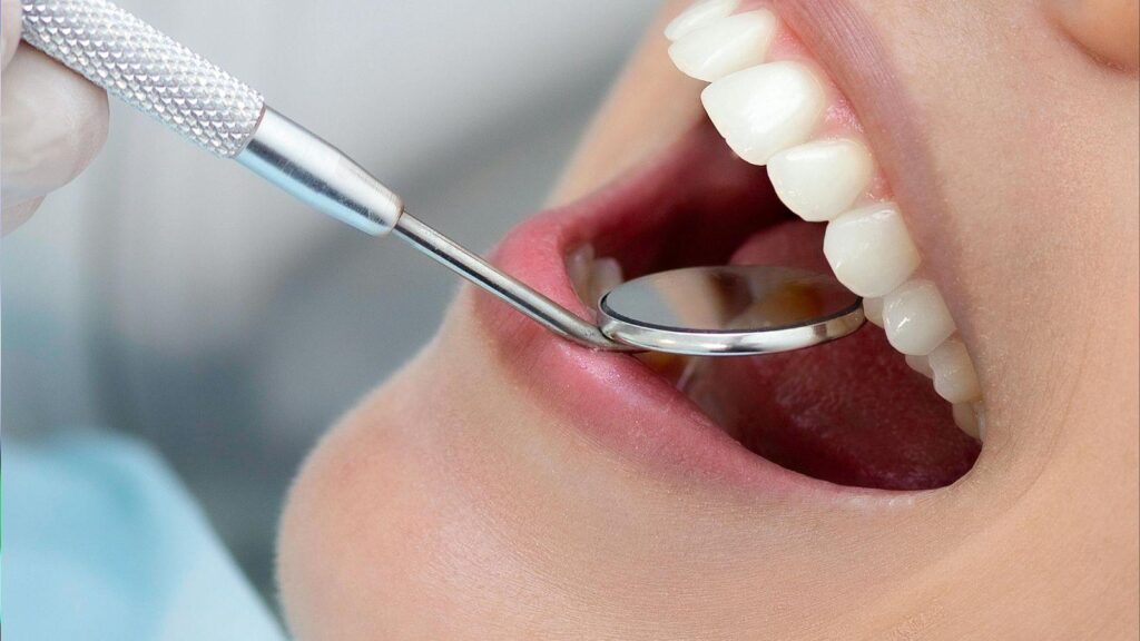
You must have adequate bone height, width, and quality where implants will be placed. Cone-beam CT (CBCT) is the primary tool to measure residual ridge dimensions, cortical thickness, and proximity to vital structures (inferior alveolar nerve, sinus floor).
If bone is insufficient, your surgeon may plan grafting (autograft, allograft, or xenograft) or staged approaches such as guided bone regeneration before implant placement.
Track radiographic signs of healing: continuous trabecular patterns, absence of radiolucency, and stable graft integration.
Clinically, assess soft-tissue coverage, keratinized gingiva presence, and any scar contracture from prior surgery that could affect flap design.
Smoking, prior radiation, and infection history alter healing expectations and may require altered timing or adjunctive therapies like hyperbaric oxygen.
Evaluating Occlusion Stability
You must have a stable, reproducible occlusion before definitive implants to avoid prosthetic overload.
Confirm orthodontic treatment is complete and that final interarch relationships do not shift after orthognathic fixation. Use mounted models or digital bite records to evaluate centric relation and canine/incisal guidance.
Assess parafunctional habits (bruxism) and existing temporomandibular joint signs; these increase risk of implant or prosthesis failure without mitigation (occlusal guards, selective occlusal adjustment).
When posterior support is lacking, consider increasing implant number, altering implant distribution, or planning screw-retained prostheses to improve load distribution.
Document occlusal scheme and prosthetic plan to guide immediate versus delayed loading decisions.
Systemic Health Factors
You need controlled systemic conditions for predictable osseointegration. Diabetes should be well controlled (HbA1c targets individualized but typically <7–8%); uncontrolled hyperglycemia raises infection and failure risk.
Review medications that affect bone metabolism or healing—bisphosphonates, denosumab, long-term corticosteroids—and coordinate with the prescribing physician for risk mitigation.
Assess prior head-and-neck radiation; irradiated bone shows higher implant failure rates and often requires pre-treatment planning such as hyperbaric oxygen or altered timelines.
Evaluate nutritional status, immune competence, and lifestyle factors (smoking, alcohol) because they directly influence soft-tissue healing and bone turnover.
Obtain informed medical clearance when major comorbidities exist and document shared decision-making about risks and alternative prosthetic options.
Treatment Timing and Planning
You need a clear schedule that aligns surgical healing, imaging, and prosthetic goals so implants are placed when bone, bite, and soft tissue are stable. Coordination among your surgeon, orthodontist, and restorative dentist determines exact dates and the sequence of steps.
Coordinating with Surgical Recovery
Plan implant placement after hard- and soft-tissue healing reaches predictable milestones. Bone remodeling and nerve recovery often require waiting 3–6 months after orthognathic osteotomies before placing implants in moved segments; shorter windows increase risk of implant failure or altered occlusion.
Monitor clinical signs: absence of infection, stable occlusion at rest and in function, and firm gingival tissues. Your surgeon will confirm radiographic bone consolidation and jaw stability. If you require bone grafting or sinus lifts, build additional healing time (typically 4–6 months) into the schedule.
Discuss staged versus simultaneous approaches. Some cases allow immediate implant placement in unchanged segments; others favor staged timing after full orthodontic decompensation and skeletal healing.
Diagnostic Imaging Approaches
Use a combination of CBCT, panoramic radiographs, and intraoral scans for implant planning after jaw surgery. CBCT gives 3D bone volume, proximity to nerves/sinuses, and assessment of osteotomy healing. Obtain a post-healing CBCT when clinical stability is evident.
Capture digital intraoral scans for occlusion, implant surgical guides, and prosthetic planning. Merge scans with CBCT to plan implant angulation, depth, and prosthetic emergence. Take repeat imaging after any secondary grafting or sinus augmentation.
Document pre- and post-surgical anatomy. Keep baseline records (pre-op CBCT and photos) to compare changes caused by orthognathic movements that affect implant site position and restorative design.
Multidisciplinary Treatment Planning
Bring your surgeon, orthodontist, and restorative dentist together for joint planning meetings. Define the final occlusal scheme, vertical dimension, and esthetic goals before implant surgery. Assign responsibilities and a timeline for appliance removal, provisional restorations, and final prostheses.
Use shared digital files (CBCT, scans, treatment plans) and coordinate chairside workflow. Resolve potential conflicts such as implant placement interfering with future orthodontic tooth movement or tooth-supported prosthesis needs. Plan temporary solutions — e.g., removable or fixed provisionals — to maintain function during healing.
Agree on contingency steps: what to do if bone is insufficient, if nerve symptoms persist, or if relapse alters occlusion. Clear decision points reduce delays and repeated surgeries.
Personalizing Timelines for Implants
Tailor timing to your age, systemic health, smoking status, and medication use. Tobacco and bisphosphonates extend healing times and raise complications; allow longer intervals or consult medical specialists. Younger patients with robust healing may shorten intervals; older or medically complex patients need longer monitoring.
Factor in orthodontic schedule and prosthetic lead times. If you need braces post-surgery, plan implant sites that won’t obstruct tooth movement, or delay implants until orthodontic goals are achieved. Build buffer time for graft incorporation, potential revisions, and custom prosthesis fabrication (typically 2–8 weeks after implant integration).
Create a written timeline with milestones (imaging, provisional, implant surgery, loading) and expected dates. Update it at each team meeting to reflect healing progress and any required changes.
Surgical Techniques and Challenges
You will face altered bone geometry, variable bone quality, and soft-tissue changes that affect implant angulation, primary stability, and esthetic outcomes. Planning, intraoperative navigation, and staged grafting commonly determine success.
Implant Placement in Altered Jaw Anatomy
Jaw repositioning, osteotomies, or fracture fixation change ridge contours and can leave you with asymmetrical bone height or redirected cortical plates. Preoperative CBCT and digital planning let you map residual bone volume, identify vital structures, and simulate implant trajectories to avoid plates, screws, or neurovascular bundles.
Use guided surgery or dynamic navigation when native anatomy is distorted to translate the plan precisely. Consider shorter, wider implants or angled implants (e.g., 30–45°) where vertical height is limited. If fixation hardware obstructs optimal placement, plan removal or place implants in a staged approach after hardware consolidation.
Managing Scar Tissue and Bone Grafts
Scarred mucosa tightness reduces flap mobility and increases dehiscence risk. You must release scars with careful incisions and, if needed, perform local tissue rearrangement or vestibuloplasty to achieve tension-free closure.
When bone is deficient, choose between particulate grafts, block grafts, or guided bone regeneration based on defect morphology. Autogenous bone offers higher integration but increases morbidity; allograft/xenograft with membranes can work for lateral defects. Stage implants after graft consolidation (4–6 months for many grafts) unless primary stability and graft type support simultaneous placement.
Optimizing Soft Tissue Integration
Peri-implant soft tissue thickness and keratinized mucosa influence long-term hygiene access and esthetics. If you lack keratinized tissue, perform soft-tissue grafting (connective tissue graft or free gingival graft) around implants to improve seal and reduce recession risk.
Use transmucosal healing abutments or provisional crowns shaped to guide tissue form and papillae. Monitor for scar-induced biotype changes and adjust prosthetic contours to minimize pressure on fragile tissues. Maintain strict plaque control and schedule frequent recalls during the first year to detect and manage mucosal complications early.
Long-Term Outcomes and Maintenance
Successful long-term results depend on predicting which implants will last, maintaining soft-tissue and bone health through daily care, and early detection of complications so you can intervene before failure.
Predicting Implant Longevity
You assess longevity by combining clinical factors and patient history. Key predictors include bone quality at the implant site, remaining native teeth status, and whether you received radiotherapy or mandibular reconstruction; irradiated bone and large reconstructive defects carry higher risk of implant loss. Implant design and surface treatments matter too—osseointegrated, roughened surfaces show better long-term stability in many cohorts.
Systemic health is crucial. Controlled diabetes and cessation of smoking improve survival odds. Prior periodontal disease increases risk for peri-implantitis, so you should treat active periodontal disease before implant placement. Track prosthetic loading: early overloading and poorly fitting restorations increase mechanical failure risk.
Oral Hygiene Strategies
You must maintain a meticulous home-care routine tailored to implants and any orthognathic changes. Clean around abutments and prosthetic margins daily with a soft-bristled brush and interdental brushes sized to the implant sulcus; avoid metal picks that can scratch implant surfaces. Use low-abrasive toothpaste and chlorhexidine rinses short-term after surgery as directed.
Professional maintenance visits should occur every 3–6 months initially. At those visits, hygienists perform semiannual debridement with nonmetallic instruments, assess soft tissue, and reinforce home care. If you have limited mouth opening after surgery, ask for specialized tools (angled brushes, single-tuft brushes) and training to reach posterior implants. Document plaque scores and peri-implant probing depths to measure progress.
Monitoring for Complications
You should receive a structured surveillance plan to detect complications early. Monitor for signs of peri-implant mucositis and peri-implantitis—redness, bleeding on probing, increasing probing depths, or suppuration—and schedule radiographs (periapical or panoramic) at baseline, one year, and then every 2–3 years or sooner if symptoms arise. Compare bone levels to baseline images to detect progressive bone loss.
Watch for prosthetic issues like screw loosening, wear of occlusal surfaces, or fracture of the restoration; adjust occlusion if you notice new mobility or discomfort. After jaw reconstruction or radiotherapy, maintain closer follow-up because complication and failure rates are higher; coordinate surveillance with your surgical team and prosthodontist for combined clinical and radiographic assessment.